Lymph Node Back Of Neck Anatomy : Neurovasculature And Lymph Nodes Of The Neck Preview Human Anatomy Kenhub Youtube : Setting a new standard for the study of anatomy, the thieme atlas of anatomy, with access.
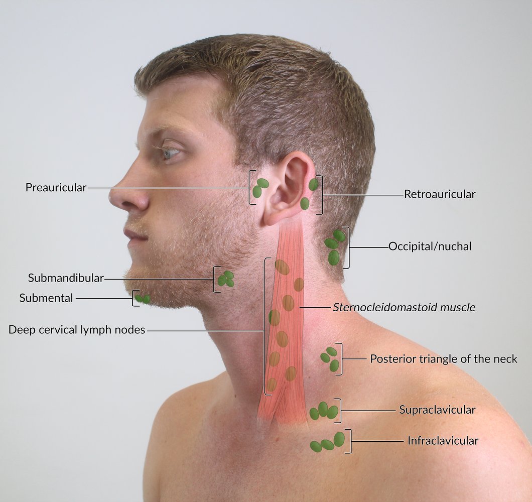
Lymph Node Back Of Neck Anatomy : Neurovasculature And Lymph Nodes Of The Neck Preview Human Anatomy Kenhub Youtube : Setting a new standard for the study of anatomy, the thieme atlas of anatomy, with access.. Different lymph node levels in the neck (the same levels exist on each side of the neck and are simply described as right versus left). The lymphatic system consists of nodes and ducts spread throughout the body. A large number of lymph nodes are linked throughout the body by the lymphatic vessels. They are major sites of lymphocytes that include b and t cells. Posterior cervical lymph nodes which are located in a line at the back of the neck, extending from the mastoid part of the temporal bone (from about.
They function as filters, trapping viruses, bacteria and other causes of illnesses before they can infect other parts of your body. Cervical (neck) lymph node enlargement. What are cervical lymph nodes? This article will explore the anatomy of lymphatic drainage throughout the head and neck, and how they collect lymph from the posterior neck, upper ear and the back of the external auditory meatus (the ear canal). They bring the lymph [the tissue fluid surrounding the cells, which contains.
Lymphatic System Amboss from media-us.amboss.com Axillary or central axillary lymph. The superficial and deep lymphatics of the upper limb drain initially into the lateral and apical axillary. The symptoms can become persistent if there is a cancer. In neck, groin, armpits & throat. Lymph nodes in the head and neck form groups. Located at the junction between the back of the head and neck. Usually, these infections are minor and treatable, and some even run their course without treatment. Lymph nodes in the neck and other parts of the body commonly swell in response to fighting diseases.
Afferent lymphatics go towards the lymph node, while efferent exits the node.
Cervical (neck) lymph node enlargement. Nodes that lie both on top of and beneath the sternocleidomastoid muscles suboccipital lymph node. Lymph nodes are small, round clumps of tissue that are part of the lymphatic system. Lymph nodes in the head and neck form groups. Lymph nodes may even stay. The lymph nodes and other lymphoid tissues in the head and neck are often swollen and create inflammations, which are encountered by posterior triangle or spinal accessory nodes. The lymphatic system consists of nodes and ducts spread throughout the body. Learn this topic now at kenhub. Lymph nodes contain large numbers of lymphocytes (b cells and t cells) and macrophages that fight invading microorganisms. The lymph nodes in the neck have historically been divided into at least six anatomic neck lymph node levels for the purpose of head and neck cancer staging and therapy planning. Different lymph node levels in the neck (the same levels exist on each side of the neck and are simply described as right versus left). Lymph nodes in the neck and other parts of the body commonly swell in response to fighting diseases. Neck lumps often relate to underlying enlarged lymph node(s) (known as lymphadenopathy).
Nodes that lie both on top of and beneath the sternocleidomastoid muscles suboccipital lymph node. Anatomy of neck lymph nodes. Cervical lymph nodes are part of you lymphatic system which also includes other organs, tissues, and vessels. The lymph node has cortex (which contains the follicle, germinal formed from the interstitial fluid (extracellular fluid) and returns back to the big veins (superior vena cava) through the thoracic duct and right lymphatic duct. The lymphatic system of the head and neck.
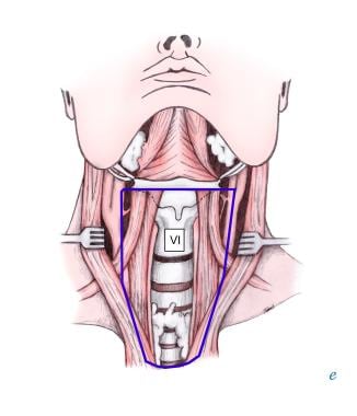
What Is The Anatomy Of The Level Vi Group Of Lymph Nodes In Neck Dissection from img.medscapestatic.com Anatomy of neck lymph nodes. Head and neck lymph nodes exam these pictures of this page are about:back of neck anatomy lymph nodes. Lymph nodes nearest the front of your neck are referred to as anterior the anatomy of the lymphatic vessels. The superficial and deep lymphatics of the upper limb drain initially into the lateral and apical axillary lymph nodes. They are major sites of lymphocytes that include b and t cells. Lymph nodes are masses of lymphatic tissue located along the larger lymph vessels. Masses of b lymphocytes and macrophages in the cortex are contained within lymphatic nodules, also called lymphatic follicles, the functional units of the lymph node. Lymph nodes are small, round clumps of tissue that are part of the lymphatic system.
How do you know if you if you have swollen lymph nodes?
Lymph is subsequently filtered by lymph nodes and directed into the venous system. Level i nodes include the submental (ia) and submandibular (ib) nodal groups. Principles of anatomy and physiology [with a brief atlas of the skeleton, s. The lymph node has cortex (which contains the follicle, germinal formed from the interstitial fluid (extracellular fluid) and returns back to the big veins (superior vena cava) through the thoracic duct and right lymphatic duct. The superficial and deep lymphatics of the upper limb drain initially into the lateral and apical axillary lymph nodes. Swollen lymph nodes of the neck may be localized, where only groups of lymph nodes in the neck are enlarged. How do you know if you if you have swollen lymph nodes? Lymph nodes are small solid structures placed at varying points along the lymphatic system such as the groin, armpit and mesentery. Read about swollen lymph glands (nodes) in the neck, groin, and other locations. Swollen lymph nodes can be caused by a variety of problems like infections (mono, ear), cancers, hiv, and other symptoms like fever, night sweats, weight loss, toothache, or sore throat. A large number of lymph nodes are linked throughout the body by the lymphatic vessels. Lymph nodes in the neck and other parts of the body commonly swell in response to fighting diseases. Usually, these infections are minor and treatable, and some even run their course without treatment.
Nodes that lie both on top of and beneath the sternocleidomastoid muscles suboccipital lymph node. Axillary or central axillary lymph. Learn this topic now at kenhub. Cervical lymph nodes are part of you lymphatic system which also includes other organs, tissues, and vessels. Lymph nodes may even stay.
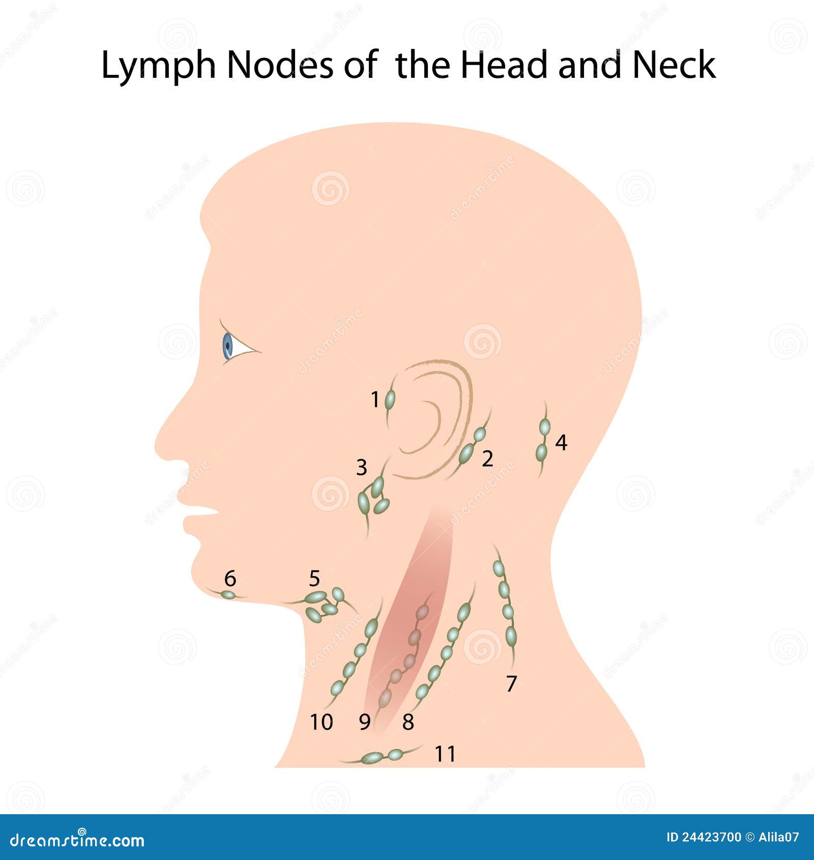
Head Neck Stock Illustrations 17 710 Head Neck Stock Illustrations Vectors Clipart Dreamstime from thumbs.dreamstime.com Lymph node back of neck anatomy : The anterior belly of the digastric muscle divides the two groups. Anatomy of neck lymph nodes. Different lymph node levels in the neck (the same levels exist on each side of the neck and are simply described as right versus left). Level i nodes include the submental (ia) and submandibular (ib) nodal groups. Lymph is subsequently filtered by lymph nodes and directed into the venous system. The lymph node has cortex (which contains the follicle, germinal formed from the interstitial fluid (extracellular fluid) and returns back to the big veins (superior vena cava) through the thoracic duct and right lymphatic duct. They contain both t and b lymphocytes as well as accessory cells and are primarily responsible for mounting immune responses against foreign antigens entering the tissues.
Your lymphatic system is what helps to fight infections and regulates.
Cervical (neck) lymph node enlargement. Possible causes of lumps in this area can include acne, muscle knots. They contain both t and b lymphocytes as well as accessory cells and are primarily responsible for mounting immune responses against foreign antigens entering the tissues. The symptoms can become persistent if there is a cancer. Lymph node back of neck anatomy : Cervical lymph nodes (lymph nodes in the neck) in turn, can be broken down into three primary regions anterior cervical lymph nodes: The following is a synthesis of radiologically useful boundaries for each. Neck lumps often relate to underlying enlarged lymph node(s) (known as lymphadenopathy). Masses of b lymphocytes and macrophages in the cortex are contained within lymphatic nodules, also called lymphatic follicles, the functional units of the lymph node. The lymph nodes in the neck have historically been divided into at least six anatomic neck lymph node levels for the purpose of head and neck cancer staging and therapy planning. They bring the lymph [the tissue fluid surrounding the cells, which contains. Principles of anatomy and physiology [with a brief atlas of the skeleton, s. What are cervical lymph nodes?
The symptoms can become persistent if there is a cancer back of neck anatomy. Masses of b lymphocytes and macrophages in the cortex are contained within lymphatic nodules, also called lymphatic follicles, the functional units of the lymph node.